Conjoint Senior Principal Scientist, NeuRA
Professor of Brain Sciences, UNSW
Director (Medicine) EPICentre, UNSW
Director (Research) NeuRA Imaging
Honorary Associate, Macquarie Centre for Cognitive Science, Macquarie University, Sydney
School of Medical Sciences, UNSW
+612 9399 1211
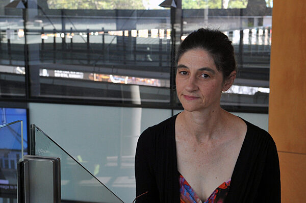
Prof Caroline Rae is a biochemist with a background in magnetic resonance and interdisciplinary brain research. She graduated with a PhD in biochemistry and NMR from The University of Sydney in 1993 and spent four years in Oxford, UK, as a Nuffield Medical Fellow where she pioneered the use of magnetic resonance spectroscopy as a tool in cognitive brain research. In 2005 she was appointed to UNSW as a New South Global Professor, one of only a handful of NHMRC R Douglas Wright Fellows subsequently appointed to chairs. She is currently director of the UNSW Node of the National Imaging Facility and holds a cross-disciplinary (STEM) appointment in medical data visualisation as a Director of the UNSW Expanded Perception and Interaction Centre (EPICentre).
The MUGgLE study is a research study on growth of muscles and tendons during childhood development. We are studying muscles of typically developing children and of children with cerebral palsy using MRI scans of the lower legs.
The MUGgLE study is a collaboration between researchers at Cerebral Palsy Alliance Research Institute, University of New South Wales and Neuroscience Research Australia (NeuRA). The study will be conducted at NeuRA’s Imaging Facility in Randwick (Sydney), Australia.
The aims of the MUGgLE study are to:
Infants aged 0 to 3 months and children aged 5 to 14 can participate in the MUGgLE study. We are looking for children with and without cerebral palsy.
Find out more on the study website: https://muggle.neura.edu.au
This project aims to better identify those at higher risk of subsequently developing Parkinson’s disease with the aim of being about to test and validate potential preventative measures. We aim to identify people at risk while they are still healthy before irreversible major cell loss has occurred.
Collaborators: Kay Double, USyd; Gabrielle Todd, UNiSA, Karl Aoun (Usyd)
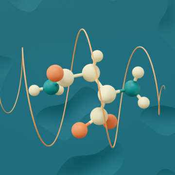
This Australian Research Council funded project (DP180101702) aims to understand the functions that amino acids play in brain, in particular in protein biosynthesis and energy generation. We are using a combination of genetic and pharmacological manipulation in conjunction with isotope labelling and NMR spectroscopy to help build our model.
Collaborators: Stefan Bröer, ANU, Brian Billups, ANU, Abhijit Das NeuRA
This study is examining the neurobiological impact of binge alcohol consumption in 16-17 yo adolescents using magnetic resonance imaging and spectroscopy along with cognitive and behavioural measures.
Collaborators: Lucette Cysique, NeuRA; Maree Teesson, NDARC Louise Mewton NDARC
Lees, B., Mewton, L., Stapinski, L, Squeglia, L. M., Rae, C. & Teesson, M. (2018) Binge drinking in young people: Protocol for a systematic review of neuropsychological, neurophysiological and neuroimaging studies. BMJ Open 8 (7) e023629.
In collaboration with colleagues in Electrical Engineering at UNSW we are developing and applying new methods for identifying and comparing brain functional and structural networks.
Main Collaborators: Victor Solo (UNSW); Ben Cassidy (NeuRA); Michael Green (NeuRA)
Cassidy, B., Rae, C. & Solo, V. (2015) Brain activity: connectivity, sparsity and mutual information. IEEE Transactions on Medical Imaging 34, 846-860.
Cassidy. B., Bowman, D., Rae, C. & Solo, V. (2018) On the reliability of individual brain activity networks. IEEE Transactions in Medical Imaging 37, 649-662.
Systems biochemistry requires the understanding and manipulation of large data sets. Here, we are applying network analysis methods from other disciplines to metabolic datasets. In collaboration with the Expanded Perception and Interaction Centre (EPICentre) at UNSW and the Australian Metabolic Phenotyping Centre based in WA, we are developing data visualisation tools for these datasets that work across a variety of 2D, and immersive platforms.
Main Collaborators: Tomasz Bednarz EPICentre; Jeremy Nicholson, Murdoch University
All cells require a constant supply of energy to survive, however the active and dynamic environment within the brain imposes not only a high basal energy requirement, but also vastly fluctuating energy demands across the course of a normal day. Fundamental to understanding brain energy metabolism is the lynchpin molecule, nicotinamide adenine dinucleotide (NAD). Despite its central importance to brain metabolism, we still do not know how the fundamental processes of NAD synthesis, recycling and catabolism are regulated in brain. Through use of highly innovative and novel strategies combining new molecular tools and flagship infrastructure, we are selectively dissecting the synthesis and roles of NAD across the 3 major classes of neural cells – neurons, astrocytes, oligodendrocytes. The results of this project will generate a systems biology platform to underpin manipulations of NAD homeostasis as well as to understand the relationship between brain NAD levels and mitochondrial function.
Main Collaborators: Gary Housley (SOMS), Matthias Klugmann (SOMS), Ben Rowlands (NeuRA)
Rowlands, B., Klugmann, M & Rae, C.D. (2017) Acetate metabolism does not reflect astrocytic activity, contributes directly to GABA synthesis, and is increased by silent information regulator 1 activation. The Journal of Neurochemistry 140, 903-918.
Sleep apnoea results in cognitive dysfunction, excessive sleepiness, doubling of workplace accidents and more than 2-fold increased motor vehicle crash risk presenting a huge health care burden in Australia and internationally. The disorder is also associated with increased risk of stroke, heart disease and possibly dementia. It is a complex disorder with multiple impacts including hypoxia, sleep deprivation, metabolic syndrome and cardiovascular risk factors.
This project aims to unravel the impact of OSA on the brain and to determine whether deleterious effects can be reversed or slowed by treatment options.
Main collaborators: Andrew Vakulin, Flinders; Ron Grunstein, Woolcock Institute, Angela D’Rozario, Woolcock Institute, Delwyn Bartlett, Woolcock Institute; Lynne Bilston, NeuRA, Michael Green, NeuRA
Miller, C.B., Rae, C.D. Green, M., Yee, B.J., Kyle, S.D., Gordon, C.J., Marshall, N.S., Espie, C.A., Grunstein, R.R. & Bartlett, D.J. (2017) An objective short-sleep insomnia disorder subtype is associated with reduced brain metabolites in vivo: a preliminary magnetic resonance spectroscopy assessment. Sleep 40, zsx148
D’Rosario, A., Bartlet, D., Wong, K.H., Sach, T, Yang, Q., Grunstein, R.R. & Rae, C.D. (2018) Brain bioenergetics during resting wakefulness are related to neurobehavioural deficits in severe obstructive sleep apnea. Sleep 41(8) zsy117.
BEN ROWLANDS PhD student
JUN CAO PhD student
ABHIJIT DAS PhD student
SALLY MCEWAN PhD student
To assess whether HIV-related brain injury is progressive in persons with suppressed HIV infection. Our study reveals covertly active or progressing HIV-related brain injury in the majority of this virally suppressed cohort, reflecting ongoing neuropathogenic processes that are only partially worsened by historical HAND and HIV duration. Longer-term studies will be important for determining the prognosis of these slowly evolving neurochemical abnormalities.
The objective of the current study was to quantify the degree of white matter (WM) abnormalities in chronic and virally suppressed HIV-infected (HIV+) persons while carefully taking into account demographic and disease factors. Diffusion tensor imaging (DTI) was conducted in 40 HIV- and 82 HIV+ men with comparable demographics and life style factors. The HIV+ sample was clinically stable with successful viral control. Diffusion was measured across 32 non-colinear directions with a b-value of 1000 s/mm; fractional anisotropy (FA) and mean diffusivity (MD) maps were quantified with Itrack IDL. Using the ENIGMA DTI protocol, FA and MD values were extracted for each participant and in 11 skeleton regions of interest (SROI) from standard labels in the JHU ICBM-81 atlas covering major striato-frontal and parietal tracks. We found no major differences in FA and MD values across the 11 SROI between study groups. Within the HIV+ sample, we found that a higher CNS penetrating antiretroviral treatment, higher current CD4+ T cell count, and immune recovery from the nadir CD4+ T cell count were associated with increased FA and decreased MD (p < 0.05-0.006), while HIV duration, symptomatic, and asymptomatic cognitive impairment were associated with decreased FA and increased MD (p < 0.01-0.004). Stability of HIV treatment and antiretroviral CNS penetration efficiency in addition to current and historical immune recovery were related to higher FA and lower MD (p = 0.04-p < 0.01). In conclusion, WM DTI measures are near normal except for patients with neurocognitive impairment and longer HIV disease duration.
To assess whether HIV-related brain injury is progressive in persons with suppressed HIV infection. Our study reveals covertly active or progressing HIV-related brain injury in the majority of this virally suppressed cohort, reflecting ongoing neuropathogenic processes that are only partially worsened by historical HAND and HIV duration. Longer-term studies will be important for determining the prognosis of these slowly evolving neurochemical abnormalities.